April 2023 Issue
ISSN 2689-291X
ISSN 2689-291X
Dextrocardia Imaging Challenges:
Dextrocardiodiagnostics!
Description
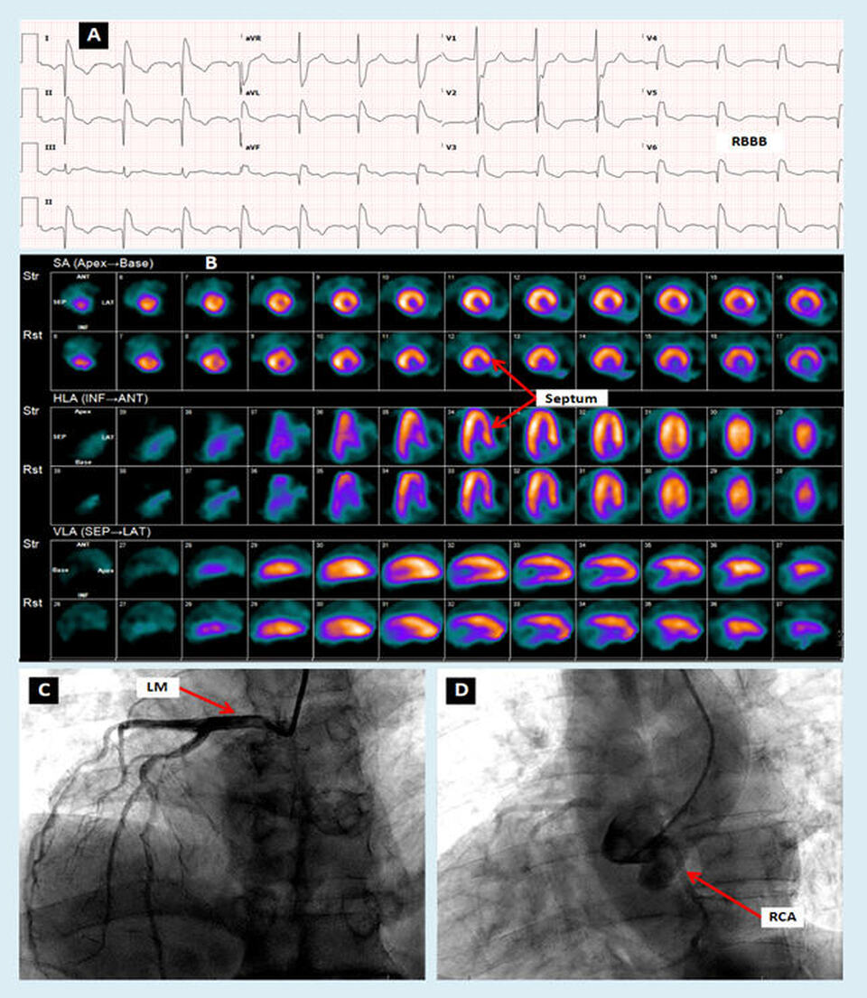
The above images illustrate some of the challenges in interpreting cardiac tests of patients with dextrocardia. Panel A shows an electrocardiogram (ECG) with right bundle branch block (RBBB) where the RSR’ pattern is inverse and is seen starting in lead V6 progressing towards lead V1. Panel B shows the nuclear results of a Lexiscan (regadenoson) Sestamibi pharmacologic stress test, with the septum appearing laterally in the short axis and horizontal long axis planes. Panel C reveals an angiogram with selective injection of the left main (LM) coronary artery which is anatomically rightward coursing towards the left ventricle. Panel D reveals a root injection with nonselective opacification of the right coronary artery (RCA) after unsuccessful attempts at engaging the right coronary ostium.
Discussion
Dextrocardia is a rare form of congenital heart disease reported in approximately 0.22% of the population [1]. It can be isolated, or associated with other congenital anomalies such as persistent right superior vena cava [2]. The association of dextrocardia with RBBB has also been reported previously [3].
When interpreting diagnostic testing in patients with dextrocardia, it is important to recognize the inverse orientation of structures. As in the above example, myocardial perfusion imaging in dextrocardia patients may lead to erroneous assumption of decreased uptake in the lateral wall if it is not recognized that what is being examined is actually the septal wall, which happens to be normally shorter with less basal perfusion [4].
Coronary angiography [5] and angioplasty [6] have been reported and can be more challenging in dextrocardia patients. The left transradial approach appears to be safe [7], and has been successfully utilized in the setting of myocardial infarction [8].
The unfamiliar right-sided (opposite) view of the coronaries can be confusing to the angiographer and can be compensated for by image double-inversion technique to adjust for the inverse and angulated views [9]. This technique has also been employed in multi-vessel percutaneous coronary intervention [10] and in primary coronary intervention in dextrocardia [11].
Conclusion
Dextrocardia is a rare congenital anomaly and presents challenges in diagnosis and treatment, mandating special approaches in imaging [12] and coronary angiography [13], often unfamiliar to practitioners. The concomitant presence of other congenital anomalies further complicates the diagnostic approach and nomenclature of such associated conditions in dextrocardia [14]. Efforts at illustrating dextrocardia using pictorial presentations are helpful in simplifying the understanding of the condition [15]. It may be worthwhile combining published innovative diagnostic approaches in patients with dextrocardia and other associated conditions in one field of dextrocardiodiagnstics.
References
Authors:
Mariam Riad, M.D.
Cardiology Fellow
University of South Alabama
Mobile, AL
Meghan Rice, B.S.
Medical Student
University of South Alabama
Mobile, AL
Bassam Omar, M.D., Ph.D.
Professor of Cardiology
University of South Alabama
Mobile, AL
The above images illustrate some of the challenges in interpreting cardiac tests of patients with dextrocardia. Panel A shows an electrocardiogram (ECG) with right bundle branch block (RBBB) where the RSR’ pattern is inverse and is seen starting in lead V6 progressing towards lead V1. Panel B shows the nuclear results of a Lexiscan (regadenoson) Sestamibi pharmacologic stress test, with the septum appearing laterally in the short axis and horizontal long axis planes. Panel C reveals an angiogram with selective injection of the left main (LM) coronary artery which is anatomically rightward coursing towards the left ventricle. Panel D reveals a root injection with nonselective opacification of the right coronary artery (RCA) after unsuccessful attempts at engaging the right coronary ostium.
Discussion
Dextrocardia is a rare form of congenital heart disease reported in approximately 0.22% of the population [1]. It can be isolated, or associated with other congenital anomalies such as persistent right superior vena cava [2]. The association of dextrocardia with RBBB has also been reported previously [3].
When interpreting diagnostic testing in patients with dextrocardia, it is important to recognize the inverse orientation of structures. As in the above example, myocardial perfusion imaging in dextrocardia patients may lead to erroneous assumption of decreased uptake in the lateral wall if it is not recognized that what is being examined is actually the septal wall, which happens to be normally shorter with less basal perfusion [4].
Coronary angiography [5] and angioplasty [6] have been reported and can be more challenging in dextrocardia patients. The left transradial approach appears to be safe [7], and has been successfully utilized in the setting of myocardial infarction [8].
The unfamiliar right-sided (opposite) view of the coronaries can be confusing to the angiographer and can be compensated for by image double-inversion technique to adjust for the inverse and angulated views [9]. This technique has also been employed in multi-vessel percutaneous coronary intervention [10] and in primary coronary intervention in dextrocardia [11].
Conclusion
Dextrocardia is a rare congenital anomaly and presents challenges in diagnosis and treatment, mandating special approaches in imaging [12] and coronary angiography [13], often unfamiliar to practitioners. The concomitant presence of other congenital anomalies further complicates the diagnostic approach and nomenclature of such associated conditions in dextrocardia [14]. Efforts at illustrating dextrocardia using pictorial presentations are helpful in simplifying the understanding of the condition [15]. It may be worthwhile combining published innovative diagnostic approaches in patients with dextrocardia and other associated conditions in one field of dextrocardiodiagnstics.
References
- Yusuf SW, Durand JB, Lenihan DJ, Swafford J. Dextrocardia: an incidental finding. Tex Heart Inst J. 2009;36(4):358-9.
- Karumbaiah K, Choe S, Ibrahim M, Omar B. Persistent right superior vena cava in a patient with dextrocardia: Case report and review of the literature. J Cardiol Cases. 2014 Jun 25;10(2):73-77.
- Adrouny ZA, Semler HJ, Griswold HE. Dextrocardia with Right Bundle Branch Block. Dis Chest. 1965 Mar;47:334-5.
- Ayeni OA, Malan N, Hammond EN, Vangu MD. Myocardial Perfusion SPECT Imaging in Dextrocardia with Situs Inversus: A Case Report. Asia Ocean J Nucl Med Biol. 2016 Summer;4(2):109-12.
- Ilia R, Gueron M. Coronary angiography in dextrocardia. Cathet Cardiovasc Diagn. 1991 Oct;24(2):150.
- Gaglani R, Gabos DK, Sangani BH. Coronary angioplasty in a patient with dextrocardia. Cathet Cardiovasc Diagn. 1989 May;17(1):45-7.
- Macdonald JE, Gardiner R, Chauhan A. Coronary angioplasty via the radial approach in an individual with dextrocardia. Int J Cardiol. 2008 Dec 17;131(1):e10-1.
- Michas G, Kaplanis I, Stougiannos P, Arapi S, Sergi E, Gavrielatos G, Trikas A. Successful transradial coronary angioplasty in a patient with dextrocardia and acute myocardial infarction. Hellenic J Cardiol. 2016 Nov-Dec;57(6):463-466.
- Goel PK. Double-inversion technique for coronary angiography viewing in dextrocardia. Catheter Cardiovasc Interv. 2005 Oct;66(2):281-5.
- Angellotti D, Esposito G, Piccolo R. Double Inversion Technique for Multivessel Primary PCI in Dextrocardia. JACC Cardiovasc Interv. 2023 Apr 10;16(7):868-869.
- Goel PK, Moorthy N. Trans-radial primary percutaneous coronary intervention in dextrocardia using double inversion technique. J Cardiol Cases. 2013 Apr 24;8(1):e31-e33.
- Maldjian PD, Saric M. Approach to dextrocardia in adults: review. AJR Am J Roentgenol. 2007 Jun;188(6 Suppl):S39-49; quiz S35-8.
- Maleki M, Tristani FE. Coronary arteriography in situs inversus dextrocardia. Chest. 1974 Feb;65(2):220-2.
- Yeo L, Luewan S, Markush D, Gill N, Romero R. Prenatal Diagnosis of Dextrocardia with Complex Congenital Heart Disease Using Fetal Intelligent Navigation Echocardiography (FINE) and a Literature Review. Fetal Diagn Ther. 2018;43(4):304-316.
- Rao PS, Rao NS. Diagnosis of Dextrocardia with a Pictorial Rendition of Terminology and Diagnosis. Children (Basel). 2022 Dec 16;9(12):1977.
Authors:
Mariam Riad, M.D.
Cardiology Fellow
University of South Alabama
Mobile, AL
Meghan Rice, B.S.
Medical Student
University of South Alabama
Mobile, AL
Bassam Omar, M.D., Ph.D.
Professor of Cardiology
University of South Alabama
Mobile, AL

