February 2022 Issue
ISSN 2689-291X
ISSN 2689-291X
Large Pericardial Cyst: Impressive & Compressive!
Description
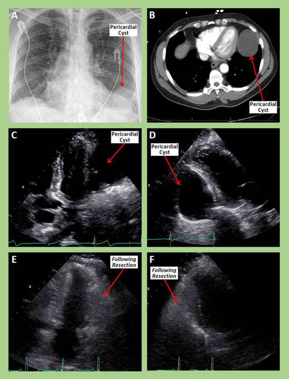
The chest roentgenogram in Figure A reveals a left lower lobe rounded shadow adjacent to the left ventricular apex, consistent with pericardial cyst. The axial computed tomographic image of the chest in Figure B better illustrates the pericardial cyst along the inferolateral and apical walls of the left ventricle and establishes its attachment to the pericardium. The 2-dimensional echocardiographic images in the apical 4 chamber view (Figure C) and modified parasternal long axis view (Figure D) demonstrate a large pericardial cyst compressing the inferolateral wall of the left ventricle. Following surgical resection, the compression is relieved as seen on the corresponding 2-D apical (Figure E) and parasternal (Figure F) images.
Discussion
Pericardial cysts are rare occurring in about 1 in 100,000 patients [1]. They are congenital anomalies caused by failure of one or more of the fetal mesenchymal lacunae to combine to form the pericardial coelom during development. The resulting localized weakness in the pericardial sac can either form a diverticulum or become a cyst upon accumulating clear fluid and losing direct communication with the pericardial sac [2].
Pericardial cysts can also occur following trauma [3], inflammatory processes such as spontaneous and post cardiothoracic surgery pericarditis [4], or in patients undergoing long-term hemodialysis [5]. About 75% of pericardial cysts are found in the right cardiophrenic angle, and close to 22% in the left cardiophrenic angle; nearly 8% are seen in the posterior or anterior/superior mediastinum [6].
An isolated pericardial cyst is usually benign [7] and often an incidental finding [8]. Most pericardial cysts (up to 75%) are asymptomatic and are discovered incidentally on routine chest imaging [9]. In symptomatic cases, symptoms are predominantly caused by mass effect, as the cyst can compress surrounding structures, such as the heart [10], the lungs and the superior vena cava [11]. Cardiac compression includes the right ventricle [12], left ventricle [13], left atrium [14] and right atrium [15]. Compression of the coronary arteries and superior vena cava has also been reported [16].
A broad range of symptoms have been reported and attributed to pericardial cysts [17]. These include chest pain [18], dyspnea [19], cough [20] and palpitations [13]. More serious presentations have been reported including obstructive shock [21], anaphylactic shock [22], cardiac tamponade [23] and respiratory failure [24].
Treatment is usually conservative; serial transthoracic echocardiograms can be performed to monitor for cyst enlargement or changes, especially in asymptomatic patients [25]. In symptomatic patients, open or minimally-invasive surgery might be indicated; percutaneous aspiration and ethanol ablation/sclerosis have also been described, although post-aspiration recurrence of the cyst has been reported to be around 33% [27, 28].
References
Authors:
Shrikar Iragavarapu, B.S.
Medical Student
University of South Alabama
Mobile, AL
Maulikkumar Patel, M.D.
Cardiology Fellow
University of South Alabama
Mobile, AL
Rajasekhar Mulyala, M.D.
Cardiology Fellow
University of South Alabama
Mobile, AL
G. Mustafa Awan, M.D.
Associate Professor of Cardiology
University of South Alabama
Mobile, AL
Christopher Malozzi, D.O.
Associate Professor of Cardiology
University of South Alabama
Mobile, AL
Bassam Omar, M.D., Ph.D.
Professor of Cardiology
University of South Alabama
Mobile, AL
The chest roentgenogram in Figure A reveals a left lower lobe rounded shadow adjacent to the left ventricular apex, consistent with pericardial cyst. The axial computed tomographic image of the chest in Figure B better illustrates the pericardial cyst along the inferolateral and apical walls of the left ventricle and establishes its attachment to the pericardium. The 2-dimensional echocardiographic images in the apical 4 chamber view (Figure C) and modified parasternal long axis view (Figure D) demonstrate a large pericardial cyst compressing the inferolateral wall of the left ventricle. Following surgical resection, the compression is relieved as seen on the corresponding 2-D apical (Figure E) and parasternal (Figure F) images.
Discussion
Pericardial cysts are rare occurring in about 1 in 100,000 patients [1]. They are congenital anomalies caused by failure of one or more of the fetal mesenchymal lacunae to combine to form the pericardial coelom during development. The resulting localized weakness in the pericardial sac can either form a diverticulum or become a cyst upon accumulating clear fluid and losing direct communication with the pericardial sac [2].
Pericardial cysts can also occur following trauma [3], inflammatory processes such as spontaneous and post cardiothoracic surgery pericarditis [4], or in patients undergoing long-term hemodialysis [5]. About 75% of pericardial cysts are found in the right cardiophrenic angle, and close to 22% in the left cardiophrenic angle; nearly 8% are seen in the posterior or anterior/superior mediastinum [6].
An isolated pericardial cyst is usually benign [7] and often an incidental finding [8]. Most pericardial cysts (up to 75%) are asymptomatic and are discovered incidentally on routine chest imaging [9]. In symptomatic cases, symptoms are predominantly caused by mass effect, as the cyst can compress surrounding structures, such as the heart [10], the lungs and the superior vena cava [11]. Cardiac compression includes the right ventricle [12], left ventricle [13], left atrium [14] and right atrium [15]. Compression of the coronary arteries and superior vena cava has also been reported [16].
A broad range of symptoms have been reported and attributed to pericardial cysts [17]. These include chest pain [18], dyspnea [19], cough [20] and palpitations [13]. More serious presentations have been reported including obstructive shock [21], anaphylactic shock [22], cardiac tamponade [23] and respiratory failure [24].
Treatment is usually conservative; serial transthoracic echocardiograms can be performed to monitor for cyst enlargement or changes, especially in asymptomatic patients [25]. In symptomatic patients, open or minimally-invasive surgery might be indicated; percutaneous aspiration and ethanol ablation/sclerosis have also been described, although post-aspiration recurrence of the cyst has been reported to be around 33% [27, 28].
References
- Kar SK, Ganguly T. Current concepts of diagnosis and management of pericardial cysts. Indian Heart J. 2017 May-Jun;69(3):364-370.
- Meredith A, Zazai IK, Kyriakopoulos C. Pericardial Cyst. 2022 Feb 13. In: StatPearls [Internet]. Treasure Island (FL): StatPearls Publishing; 2022 Jan–.
- Saldaña Dueñas C, Hernández Galán A. Posttraumatic pericardial cyst. An Sist Sanit Navar. 2015 Sep-Dec;38(3):475-8.
- Imran TF, Shah R, Qavi AH, Waller A, Kim B. Pleuropericarditis complicated by a pericardial cyst. J Cardiol Cases. 2015 Jul 27;12(5):156-158.
- Pugliatti P, Donato R, Crea P, Zito C, Patanè S. Image Diagnosis: Pericardial Cyst in a Dialysis Patient. J Cardiovasc Ultrasound. 2016 Jun;24(2):177-8.
- Sokouti M, Halimi M, Golzari SE. Pericardial cyst presented as chronic cough: a rare case report. Tanaffos. 2012;11(4):60-2.
- Nayak K, Shetty RK, Vivek G, Pai UM. Pericardial cyst: a benign anomaly. BMJ Case Rep. 2012 Sep 4;2012:bcr0320125984.
- Lin AN, Lin S, Lin K, Raju F. Pericardial incidentaloma: benign pericardial cyst. BMJ Case Rep. 2017 May 15;2017:bcr2017220097.
- Khayata M, Alkharabsheh S, Shah NP, Klein AL. Pericardial Cysts: a Contemporary Comprehensive Review. Curr Cardiol Rep. 2019 May 30;21(7):64.
- Islas F, de Agustin JA, Gomez de Diego JJ, Olmos C, Ferrera C, Luaces M, Cabeza B, Macaya C, Pérez de Isla L. Giant pericardial cyst compressing the heart. J Am Coll Cardiol. 2013 Sep 3;62(10):e19.
- Kaul P, Javangula K, Farook SA. Massive benign pericardial cyst presenting with simultaneous superior vena cava and middle lobe syndromes. J Cardiothorac Surg. 2008 May 21;3:32.
- Mwita JC, Chipeta P, Mutagaywa R, Rugwizangoga B, Ussiri E. Pericardial cyst with right ventricular compression. Pan Afr Med J. 2012;12:60.
- Noori NM, Shafighi Shahri E, Soleimanzadeh Mousavi SH. Large Congenital Pericardial Cyst Presented by Palpitation and Left Ventricle Posterior Wall Compression: A Rare Case Report. Pediatr Rep. 2021 Jan 15;13(1):57-64.
- Seo GW, Seol SH, Jeong HJ, Seo MG, Song PS, Kim DK, Kim KH, Kim DI, Kang MJ, Kim JY. A large pericardial cyst compressing the left atrium presenting as a pericardiopleural efussion. Heart Lung Circ. 2014 Dec;23(12):e273-5.
- Martins IM, Fernandes JM, Gelape CL, Braulio R, Silva Vde C, Nunes Mdo C. A large pericardial cyst presenting with compression of the right-side cardiac chambers. Rev Bras Cir Cardiovasc. 2011 Jul-Sep;26(3):504-7.
- Parsons C, Zhao CB, Huang J. Gigantic Pericardial Bronchogenic Cyst Compressing Superior Vena Cava and Coronary Artery. Anesthesiology. 2019 Sep;131(3):667.
- Najib MQ, Chaliki HP, Raizada A, Ganji JL, Panse PM, Click RL. Symptomatic pericardial cyst: a case series. Eur J Echocardiogr. 2011 Nov;12(11):E43.
- Varvarousis D, Tampakis K, Dremetsikas K, Konstantinedes P, Mantas I. Pericardial cyst: An unusual cause of chest pain. J Cardiol Cases. 2015 Jul 3;12(4):130-132.
- Caramori JE, Miozzo L, Formigheri M, Barcellos C, Grando M, Trentin T. Dispnéia por compressão de estruturas mediastinais por cisto pericárdico [Dyspnea through compression of mediastinal structures due to pericardial cyst]. Arq Bras Cardiol. 2005 Jun;84(6):486-7. Portuguese.
- Makar M, Makar G, Yousef K. Large Pericardial Cyst Presenting as Acute Cough: A Rare Case Report. Case Rep Cardiol. 2018 Dec 5;2018:4796903.
- Lee I, Parikh N, Sane D. A Pericardial Cyst Causing Obstructive Shock: A Case Report. JACC March 12, 2019 Volume 73, Issue 9.
- Cimpoesu D, Stoica L, Paulet A, MD, Petris A. A Case of Anaphylactic Shock Due to Pericardial Hydatid Cyst. Chest. 2012;142(4_MeetingAbstracts):322A.
- Bandeira FC, de Sá VP, Moriguti JC, Rodrigues AJ, Jurca MC, Almeida-Filho OC, Marin-Neto JA, Maciel BC. Cardiac tamponade: an unusual complication of pericardial cyst. J Am Soc Echocardiogr. 1996 Jan-Feb;9(1):108-12.
- Dassan M, Saxena A, Seneviratne C, Kupfer Y, Brichkov I, Irukulla P, Patel J. Respiratory Failure Caused by Large Pericardial Cyst. Chest. 2015;148(4_MeetingAbstracts):287A.
- Alkharabsheh S, Gentry Iii JL, Khayata M, Gupta N, Schoenhagen P, Flamm S, Murthy S, Klein AL. Clinical Features, Natural History, and Management of Pericardial Cysts. Am J Cardiol. 2019 Jan 1;123(1):159-163.
- Weder W, Klotz HP, von Segesser L, Largiadèr F. Thoracoscopic resection of a pericardial cyst: a case report. J Thorac Cardiovasc Surg. 1994 Jan;107(1):313-4.
- Sharma R, Harden S, Peebles C, Dawkins KD. Percutaneous aspiration of a pericardial cyst: an acceptable treatment for a rare disorder. Heart. 2007 Jan;93(1):22.
- Kinoshita Y, Shimada T, Murakami Y, Sano K, Tanabe K, Ishinaga Y, Kato H, Murakami R, Morioka S. Ethanol sclerosis can be a safe and useful treatment for pericardial cyst. Clin Cardiol. 1996 Oct;19(10):833-5.
Authors:
Shrikar Iragavarapu, B.S.
Medical Student
University of South Alabama
Mobile, AL
Maulikkumar Patel, M.D.
Cardiology Fellow
University of South Alabama
Mobile, AL
Rajasekhar Mulyala, M.D.
Cardiology Fellow
University of South Alabama
Mobile, AL
G. Mustafa Awan, M.D.
Associate Professor of Cardiology
University of South Alabama
Mobile, AL
Christopher Malozzi, D.O.
Associate Professor of Cardiology
University of South Alabama
Mobile, AL
Bassam Omar, M.D., Ph.D.
Professor of Cardiology
University of South Alabama
Mobile, AL

