January 2022 Issue
ISSN 2689-291X
ISSN 2689-291X
Persistent Left Superior Vena Cava:
A Left-Sided Short Circuit!
Description
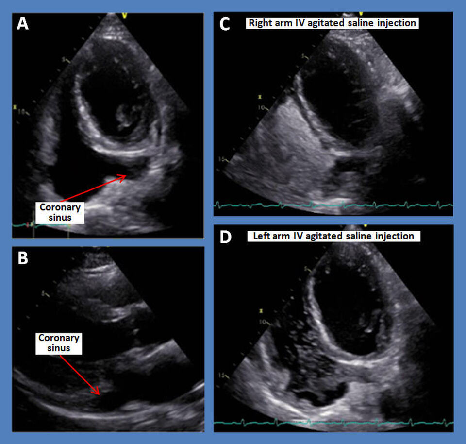
The above echocardiographic images reveal a dilated coronary sinus in a modified apical 4-chamber view (Fig. A) and parasternal long axis view (Fig. B). Injection of IV agitated saline into the right arm (Fig. C) routes the bubbles into the right superior vena cava inserting into the right arm with an expected appearance of the bubbles into the right atrium without opacification of the coronary sinus. However, injection of the agitated saline into the left arm (Fig. D) results in an unexpected initial appearance of the bubbles into the dilated coronary sinus pouring into the right atrium, indicating the presence of a persistent superior vena cava inserting into the coronary sinus.
Discussion
A left superior vena cava is a venous structure that exists during embryological development and usually disappears after birth [1]. However, some individuals have a persistent left superior vena cava, which is considered the most common thoracic venous anomaly, with a prevalence of 0.3 – 0.5% in the general population [2]. For most patients, this anomaly does not cause any hemodynamic compromise and is completely asymptomatic [3].
Embryonic vessels includes two superior cardinal veins which drain blood from the cranial portion of the fetus and an inferior caudal vein which drains the caudal portion [4]. Around the eighth week of gestation the left and right superior cardinal veins form an anastomosis which eventually forms the brachiocephalic vein. The caudal portion of the right superior cardinal vein forms the superior vena cava while the caudal portion of the left regresses to form the ligament of Marshall. If the caudal portion of the left superior cardinal vein does not regress, a vein that drains into the coronary sinuses forms, resulting in a persistent left superior vena cava. Variations of this anomaly could include a right and left superior vena cava with an Innominate vein bridging or the right superior vena cava could regress leading to a solitary left superior vena cava [5]. Most patients with this deformity have a left SVC that drains into the right atrium via the coronary sinuses. Some cases the left SVC drain into the left atrium which causes a shunt and potential paradoxical embolism [6, 7].
Most cases are diagnosed incidentally on cardiac imaging [8]. If a Swan-Ganz catheter is being placed via left subclavian, it will pass through the left superior vena cava and into the coronary sinus [9]. More definitive diagnosis is established via echocardiogram with bubble study [10]. TTE will show a dilated coronary sinus and a bubble study must be performed on both upper extremities as shown above. When agitated saline is administered via the right upper extremity it will be seen in the right atrium first. However, when administered via the left upper extremity, it will be seen first in the coronary sinus, then in the right atrium. Further imaging can be obtained via multislice CT or MR venography [11].
Most complications arise during procedures that require vascular access. The vasculature on the left can complicate placement of Swan-Ganz catheters, central lines, and ICDs [12]. Other complications associated with accessing the left SVC include arrhythmias [13], tamponade, cardiogenic shock [14], and coronary sinus thrombosis [15]. These complications are rare and are becoming less common with modern technology. In cardiac surgery, the malformation can be a contraindication to retrograde cardioplegia, requiring the coronary sinus to be dissected to permit reanastamosis of left SVC to the right atrium [16].
References
Authors:
Alexis Parks, D.O.
Internal Medicine Resident
University of South Alabama
Mobile, AL
Nikky Bardia, M.D.
Cardiology Fellow
University of South Alabama
Mobile, AL
Christopher Malozzi, D.O.
Associate Professor of Cardiology
University of South Alabama
Mobile, AL
Bassam Omar, M.D., Ph.D.
Professor of Cardiology
University of South Alabama
Mobile, AL
The above echocardiographic images reveal a dilated coronary sinus in a modified apical 4-chamber view (Fig. A) and parasternal long axis view (Fig. B). Injection of IV agitated saline into the right arm (Fig. C) routes the bubbles into the right superior vena cava inserting into the right arm with an expected appearance of the bubbles into the right atrium without opacification of the coronary sinus. However, injection of the agitated saline into the left arm (Fig. D) results in an unexpected initial appearance of the bubbles into the dilated coronary sinus pouring into the right atrium, indicating the presence of a persistent superior vena cava inserting into the coronary sinus.
Discussion
A left superior vena cava is a venous structure that exists during embryological development and usually disappears after birth [1]. However, some individuals have a persistent left superior vena cava, which is considered the most common thoracic venous anomaly, with a prevalence of 0.3 – 0.5% in the general population [2]. For most patients, this anomaly does not cause any hemodynamic compromise and is completely asymptomatic [3].
Embryonic vessels includes two superior cardinal veins which drain blood from the cranial portion of the fetus and an inferior caudal vein which drains the caudal portion [4]. Around the eighth week of gestation the left and right superior cardinal veins form an anastomosis which eventually forms the brachiocephalic vein. The caudal portion of the right superior cardinal vein forms the superior vena cava while the caudal portion of the left regresses to form the ligament of Marshall. If the caudal portion of the left superior cardinal vein does not regress, a vein that drains into the coronary sinuses forms, resulting in a persistent left superior vena cava. Variations of this anomaly could include a right and left superior vena cava with an Innominate vein bridging or the right superior vena cava could regress leading to a solitary left superior vena cava [5]. Most patients with this deformity have a left SVC that drains into the right atrium via the coronary sinuses. Some cases the left SVC drain into the left atrium which causes a shunt and potential paradoxical embolism [6, 7].
Most cases are diagnosed incidentally on cardiac imaging [8]. If a Swan-Ganz catheter is being placed via left subclavian, it will pass through the left superior vena cava and into the coronary sinus [9]. More definitive diagnosis is established via echocardiogram with bubble study [10]. TTE will show a dilated coronary sinus and a bubble study must be performed on both upper extremities as shown above. When agitated saline is administered via the right upper extremity it will be seen in the right atrium first. However, when administered via the left upper extremity, it will be seen first in the coronary sinus, then in the right atrium. Further imaging can be obtained via multislice CT or MR venography [11].
Most complications arise during procedures that require vascular access. The vasculature on the left can complicate placement of Swan-Ganz catheters, central lines, and ICDs [12]. Other complications associated with accessing the left SVC include arrhythmias [13], tamponade, cardiogenic shock [14], and coronary sinus thrombosis [15]. These complications are rare and are becoming less common with modern technology. In cardiac surgery, the malformation can be a contraindication to retrograde cardioplegia, requiring the coronary sinus to be dissected to permit reanastamosis of left SVC to the right atrium [16].
References
- Sondermeijer BM, Macgillavry MR, Tan HL. Left superior vena cava, a remnant of embryological development. Neth Heart J. 2008 May;16(5):173-4.
- Tyrak KW, Holda J, Holda MK, Koziej M, Piatek K, Klimek-Piotrowska W. Persistent left superior vena cava. Cardiovasc J Afr. 2017 May 23;28(3):e1-e4.
- Goyal SK, Punnam SR, Verma G, Ruberg FL. Persistent left superior vena cava: a case report and review of literature. Cardiovasc Ultrasound. 2008 Oct 10;6:50.
- Ghandour A, Karuppasamy K, Rajiah P. Congenital Anomalies of the Superior Vena Cava: Embryological Correlation, Imaging Perspectives, and Clinical Relevance. Can Assoc Radiol J. 2017 Nov;68(4):456-462.
- Uçar O, Paşaoğlu L, Ciçekçioğlu H, Vural M, Kocaoğlu I, Aydoğdu S. Persistent left superior vena cava with absent right superior vena cava: a case report and review of the literature. Cardiovasc J Afr. 2010 May-Jun;21(3):164-6.
- Gupta R, Pearson A. Diagnosis of persistent left superior vena cava draining directly into the left atrium. N Am J Med Sci. 2013 Aug;5(8):496-7.
- Hutyra M, Skala T, Sanak D, Novotny J, Köcher M, Taborsky M. Persistent left superior vena cava connected through the left upper pulmonary vein to the left atrium: an unusual pathway for paradoxical embolization and a rare cause of recurrent transient ischaemic attack. Eur J Echocardiogr. 2010 Oct;11(9):E35.
- Morgan LG, Gardner J, Calkins J. The incidental finding of a persistent left superior vena cava: implications for primary care providers-case and review. Case Rep Med. 2015;2015:198754.
- Huang YL, Wu MT, Pan HB, Yang CF. Aberrant course of Swan-Ganz catheter revealing persistent left superior vena cava. Zhonghua Yi Xue Za Zhi (Taipei). 2002 Aug;65(8):403-6.
- Pardinas Gutierrez MA, Escobar LA, Blumer V, Cabrera JL. Incidental finding of persistent left superior vena cava after 'bubble study' verification of central venous catheter. BMJ Case Rep. 2017 Jul 31;2017:bcr2017220133.
- Sonavane SK, Milner DM, Singh SP, Abdel Aal AK, Shahir KS, Chaturvedi A. Comprehensive Imaging Review of the Superior Vena Cava. Radiographics. 2015 Nov-Dec;35(7):1873-92.
- Zhou Q, Murthy S, Pattison A, Werder G. Central venous access through a persistent left superior vena cava: a case series. J Vasc Access. 2016 Sep 21;17(5):e143-7.
- Corîci OM, Gașpar M, Mornoș A, Iancău M. Cardiac Arrhythmias in Patient with Isolated Persistent Left Superior Vena Cava. Curr Health Sci J. 2017 Apr-Jun;43(2):163-166.
- Wissner E, Tilz R, Konstantinidou M, Metzner A, Schmidt B, Chun KR, Kuck KH, Ouyang F. Catheter ablation of atrial fibrillation in patients with persistent left superior vena cava is associated with major intraprocedural complications. Heart Rhythm. 2010 Dec;7(12):1755-60.
- Moey MYY, Ebin E, Marcu CB. Venous varices of the heart: a case report of spontaneous coronary sinus thrombosis with persistent left superior vena cava. Eur Heart J Case Rep. 2018 Aug 16;2(3):yty092.
- Fernando RJ, Johnson SD. Inability to Utilize Retrograde Cardioplegia due to a Persistent Left Superior Vena Cava. Case Rep Anesthesiol. 2017;2017:4671856.
Authors:
Alexis Parks, D.O.
Internal Medicine Resident
University of South Alabama
Mobile, AL
Nikky Bardia, M.D.
Cardiology Fellow
University of South Alabama
Mobile, AL
Christopher Malozzi, D.O.
Associate Professor of Cardiology
University of South Alabama
Mobile, AL
Bassam Omar, M.D., Ph.D.
Professor of Cardiology
University of South Alabama
Mobile, AL

