January 2019 Issue
ISSN 2689-291X
ISSN 2689-291X
Challenging Images
Cardiac Myxoma.. So Common, Yet So Rare!
Description
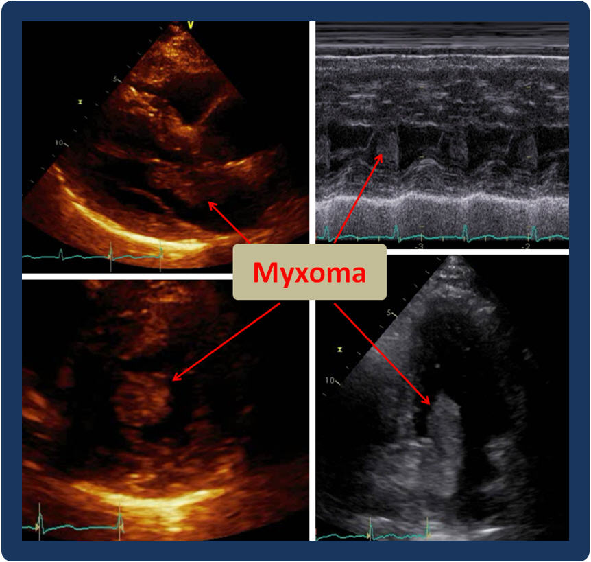
Cardiac myxoma is a benign tumor of the heart that can be responsible for a variety of systemic, inflammatory and embolic manifestations [1]. Echocardiography usually provides the initial suspicion or diagnosis, however, multimodality imaging is often required for more accurate definition [2]. Myxoma is often sporadic and isolated, but may also be multiple and recurrent, part of the Carney complex, an autosomal dominant syndrome associated with spotty skin pigmentation, endocrinopathy, in addition to endocrine and nonendocrine tumors [3]. Surgical excision is often curative [4], with long term follow-up needed to assess for recurrence [5].
When discussing cardiac myxoma in relation to other cardiac tumors, it is a celebrity, often described as the most common (> 50%) benign or primary cardiac tumor [6, 7]. However, cardiac tumors overall are uncommon, and therefore, cardiac myxoma is often reported as a rare finding, mostly in case reports or case series [8, 9].
References:
Authors:
Sarina Sachdev, M.D.
Cardiology Fellow
University of South Alabama
Mobile, AL
Landai Nguyen, D.O.
Cardiology Fellow
University of South Alabama
Mobile, AL
Hassan Tahir, M.D.
Cardiology Fellow
University of South Alabama
Mobile, AL
Farnoosh Rahimi, M.D.
Assistant Professor of Cardiology
University of South Alabama
Mobile, AL
Barbara Burckhartt, M.D.
Associate Professor of Cardiology
University of South Alabama
Mobile, AL
Christopher Malozzi, D.O.
Assistant Professor of Cardiology
University of South Alabama
Mobile, AL
Bassam Omar, M.D., Ph.D.
Professor of Cardiology
University of South Alabama
Mobile, AL
Cardiac myxoma is a benign tumor of the heart that can be responsible for a variety of systemic, inflammatory and embolic manifestations [1]. Echocardiography usually provides the initial suspicion or diagnosis, however, multimodality imaging is often required for more accurate definition [2]. Myxoma is often sporadic and isolated, but may also be multiple and recurrent, part of the Carney complex, an autosomal dominant syndrome associated with spotty skin pigmentation, endocrinopathy, in addition to endocrine and nonendocrine tumors [3]. Surgical excision is often curative [4], with long term follow-up needed to assess for recurrence [5].
When discussing cardiac myxoma in relation to other cardiac tumors, it is a celebrity, often described as the most common (> 50%) benign or primary cardiac tumor [6, 7]. However, cardiac tumors overall are uncommon, and therefore, cardiac myxoma is often reported as a rare finding, mostly in case reports or case series [8, 9].
References:
- Jain S, Maleszewski JJ, Stephenson CR, et al. Current diagnosis and management of cardiac myxomas. Expert Rev Cardiovasc Ther. 2015 Apr;13(4):369-75.
- Colin GC, Gerber BL, Amzulescu M, et al. Cardiac myxoma: a contemporary multimodality imaging review. Int J Cardiovasc Imaging. 2018 Nov;34(11):1789-1808.
- Tamura Y, Seki T. Carney complex with right ventricular myxoma following second excision of left atrial myxoma. Ann Thorac Cardiovasc Surg. 2014;20 Suppl:882-4.
- Lee KS, Kim GS, Jung Y, et al. Surgical resection of cardiac myxoma-a 30-year single institutional experience. J Cardiothorac Surg. 2017 Mar 27;12(1):18.
- Kuroczyński W, Peivandi AA, Ewald P, et al. Cardiac myxomas: short- and long-term follow-up. Cardiol J. 2009;16(5):447-54.
- Rodriguez Ziccardi M, Limaiem F. Cancer, Cardiac. StatPearls [Internet]. Treasure Island (FL): StatPearls Publishing; 2018-2019 Jan 20.
- Khan MA, Khan AA, Waseem M. Surgical experience with cardiac myxomas. J Ayub Med Coll Abbottabad. 2008 Apr-Jun;20(2):76-9.
- Ermek T, Aybek N, Zhang WM, et al. A rare case of biventricular myxoma. J Cardiothorac Surg. 2017 Mar 27;12(1):17.
- Wang H, Li Q, Xue M, et al. Cardiac Myxoma: A Rare Case Series of 3 Patients and a Literature Review. J Ultrasound Med. 2017 Nov;36(11):2361-2366.
Authors:
Sarina Sachdev, M.D.
Cardiology Fellow
University of South Alabama
Mobile, AL
Landai Nguyen, D.O.
Cardiology Fellow
University of South Alabama
Mobile, AL
Hassan Tahir, M.D.
Cardiology Fellow
University of South Alabama
Mobile, AL
Farnoosh Rahimi, M.D.
Assistant Professor of Cardiology
University of South Alabama
Mobile, AL
Barbara Burckhartt, M.D.
Associate Professor of Cardiology
University of South Alabama
Mobile, AL
Christopher Malozzi, D.O.
Assistant Professor of Cardiology
University of South Alabama
Mobile, AL
Bassam Omar, M.D., Ph.D.
Professor of Cardiology
University of South Alabama
Mobile, AL
Challenging Images
Cardiac CAT.. Where Does it Like to Hide?
Description
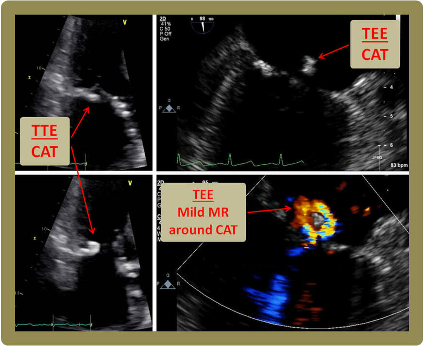
Cardiac calcified amorphous tumor (CAT) is a poorly characterized nonneoplastic endocardially based intracavitary cardiac masses, first describe as such by Reynolds and colleagues [1]. It has been reported more often in patients with end-stage renal disease and on hemodialysis, likely due to ectopic calcifications [2, 3]. Cardiac CAT can be and incidental finding on an imaging study [4], but may also present with detrimental embolic manifestations [5]. Although more often they are initially diagnosed on echocardiography, multimodality imaging may provide better delineation and localization [6]. Depending on its embolization potential or disruption of valvular competency, treatment can be conservative or surgical. As in the image above, Cardiac CAT can present as a mass swinging from the mitral valve or annulus, and must be differentiated from endocarditis [7, 8].
Although it has been reported and removed from any cardiac chamber, cardiac CAT appears to have a predilection to hiding along the mitral valve annulus, especially in dialysis patients [9]. It should, therefore, be carefully differentiated from heavy mitral annular calcifications [10].
References:
Authors:
Muhammad Awan, M.D.
Cardiology Fellow
University of South Alabama
Mobile, AL
Sajjad Ahmad, M.D.
Cardiology Fellow
University of South Alabama
Mobile, AL
Muhammad Rafique, M.D.
Cardiology Fellow
University of South Alabama
Mobile, AL
Sarina Sachdev, M.D.
Cardiology Fellow
University of South Alabama
Mobile, AL
Landai Nguyen, D.O.
Cardiology Fellow
University of South Alabama
Mobile, AL
Hassan Tahir, M.D.
Cardiology Fellow
University of South Alabama
Mobile, AL
Barbara Burckhartt, M.D.
Associate Professor of Cardiology
University of South Alabama
Mobile, AL
Bassam Omar, M.D., Ph.D.
Professor of Cardiology
University of South Alabama
Mobile, AL
Cardiac calcified amorphous tumor (CAT) is a poorly characterized nonneoplastic endocardially based intracavitary cardiac masses, first describe as such by Reynolds and colleagues [1]. It has been reported more often in patients with end-stage renal disease and on hemodialysis, likely due to ectopic calcifications [2, 3]. Cardiac CAT can be and incidental finding on an imaging study [4], but may also present with detrimental embolic manifestations [5]. Although more often they are initially diagnosed on echocardiography, multimodality imaging may provide better delineation and localization [6]. Depending on its embolization potential or disruption of valvular competency, treatment can be conservative or surgical. As in the image above, Cardiac CAT can present as a mass swinging from the mitral valve or annulus, and must be differentiated from endocarditis [7, 8].
Although it has been reported and removed from any cardiac chamber, cardiac CAT appears to have a predilection to hiding along the mitral valve annulus, especially in dialysis patients [9]. It should, therefore, be carefully differentiated from heavy mitral annular calcifications [10].
References:
- Reynolds C, Tazelaar HD, Edwards WD. Calcified amorphous tumor of the heart (cardiac CAT). Hum Pathol. 1997 May;28(5):601-6.
- Yoshimura S, Kawano H1, Minami T, Tsuneto A, et al. Cardiac Calcified Amorphous Tumors in a Patient with Hemodialysis for Diabetic Nephropathy. Intern Med. 2017 Nov 15;56(22):3057-3060.
- Takeuchi T, Dohi K, Sato Y, et al. Calcified amorphous tumor of the heart in a hemodialysis patient. Echocardiography. 2016 Dec;33(12):1926-1928.
- Kawata T, Konishi H, Amano A, et al. Wavering calcified amorphous tumour of the heart in a haemodialysis patient. Interact Cardiovasc Thorac Surg. 2013 Feb;16(2):219-20.
- Kyaw K, Latt H, Aung SSM, et al. A Case of Cardiac Calcified Amorphous Tumor Presenting with Concomitant ST-ElevationMyocardial Infarction and Occipital Stroke and a Brief Review of the Literature. Case Rep Cardiol. 2017;2017:8578031.
- Xu F, Xiao Z, Peng L, et al. A Rare Case of Cardiac Calcified Amorphous Tumor: Multi-Modality Imaging Evaluation. Am J Case Rep. 2018 Feb 27;19:214-217.
- de Hemptinne Q, Bar JP, de Cannière D, et al. Swinging cardiac calcified amorphous tumour arising from a calcified mitral annulus in a patientwith normal renal function. BMJ Case Rep. 2015 Jan 7;2015. pii: bcr2014207401.
- Fujiwara M, Watanabe H, Iino T, et al. Two cases of calcified amorphous tumor mimicking mitral valve vegetation. Circulation. 2012 Mar 13;125(10):e432-4.
- de Hemptinne Q, de Cannière D, Vandenbossche JL, et al. Cardiac calcified amorphous tumor: A systematic review of the literature. Int J Cardiol Heart Vasc. 2015 Jan 29;7:1-5.
- Nakamaru R, Oe H, Iwakura K, et al. Calcified amorphous tumor of the heart with mitral annular calcification: a case report. J Med Case Rep. 2017 Jul 18;11(1):195
Authors:
Muhammad Awan, M.D.
Cardiology Fellow
University of South Alabama
Mobile, AL
Sajjad Ahmad, M.D.
Cardiology Fellow
University of South Alabama
Mobile, AL
Muhammad Rafique, M.D.
Cardiology Fellow
University of South Alabama
Mobile, AL
Sarina Sachdev, M.D.
Cardiology Fellow
University of South Alabama
Mobile, AL
Landai Nguyen, D.O.
Cardiology Fellow
University of South Alabama
Mobile, AL
Hassan Tahir, M.D.
Cardiology Fellow
University of South Alabama
Mobile, AL
Barbara Burckhartt, M.D.
Associate Professor of Cardiology
University of South Alabama
Mobile, AL
Bassam Omar, M.D., Ph.D.
Professor of Cardiology
University of South Alabama
Mobile, AL


