October 2023 Issue
ISSN 2689-291X
ISSN 2689-291X
Mid Left Ventricular Takotsubo:
A Stress Cardiomyopathy Variant!
Description
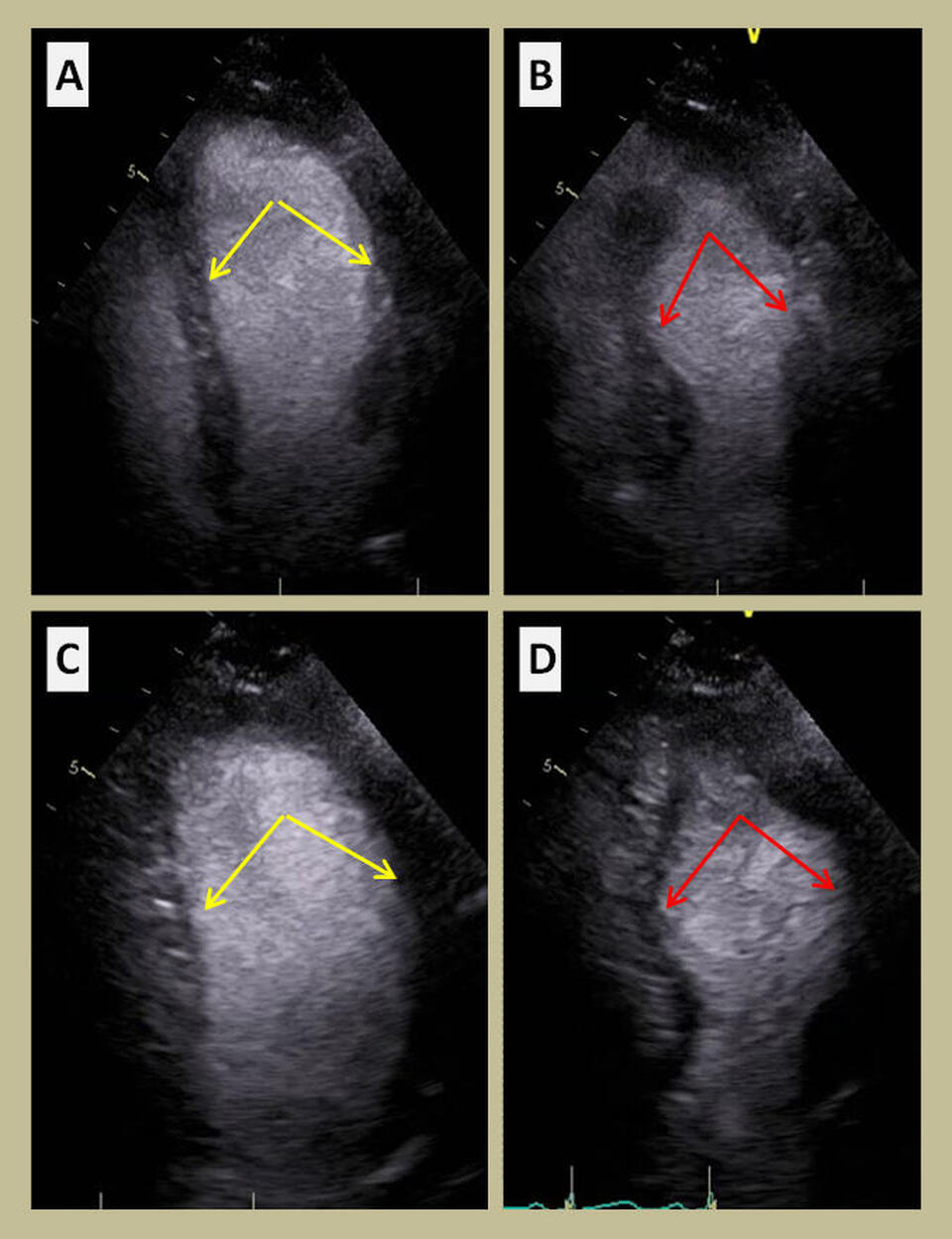
The above transthoracic echocardiogram images demonstrate 2-dimensional (2D) apical 4-chamber view frames in end-diastole (A) and end-systole (B) revealing bulging out (dyskinesis) of the mid anterolateral and inferoseptal wall segments of the left ventricular (LV) cavity. The 2-D apical 2-chamber views in diastole (C) and systole (D) demonstrate a similar mid LV cavity bulging of the anterior and inferior wall segments. The exclusive wall motion abnormality of the middle LV segments, sparing the basal and apical segments, is a rare variant of stress cardiomyopathy (Takotsubo).
Discussion
Takotsubo cardiomyopathy was first reported in Japan in the 1990s and initially attributed to multivessel vasospasm [1]. It is defined as temporary wall motion abnormality of the left ventricle in the absence of obstructive coronary artery disease correlating with such wall motion abnormality [2]. It is thought to be caused by enhanced sympathetic stimulation resulting in potential plaque rupture, multi-vessel spasm of the coronary arteries, direct myocyte catecholamine cardiotoxicity, and dysfunction of the microcirculation [3]. It is predominantly found in postmenopausal women and presents after significant emotional or physical stressors [4]. Patients can present with acute coronary syndrome signs and symptoms, elevated troponin, and ST-elevation on EKG, especially in atypical variants [5].
Takotsubo cardiomyopathy can be widely classified as a typical variant which is more common and has apical ballooning of the LV during systole with hyperkinesis of basal segments, mimicking acute myocardial infarction [6]. However, other atypical variants also exist such as basal, focal, mid-ventricular, biventricular, isolated right ventricular and global Takotsubo cardiomyopathy [7]. Mid-ventricular Takotsubo cardiomyopathy is a rare (~15%) and reversible myocardial injury presenting with distinctive regional wall motion abnormalities of the mid LV segments [8]. Rare occurrence of different Takotsubo ballooning patterns in the same patient has been reported [9]. Diagnostic workup of Takotsubo syndrome based on clinical presentation, biomarkers, imaging and calculation of an InterTAK diagnostic score has been suggested [10].
References
Authors:
Sanchitha Nagaraj, M.D.
Visiting Resident
University of South Alabama
Mobile, AL
Nupur Shah, M.D.
Cardiology Fellow
University of South Alabama
Mobile, AL
Celestine Odigwe, M.D.
Cardiology Fellow
University of South Alabama
Mobile, AL
Christopher Malozzi, D.O.
Associate Professor of Cardiology
University of South Alabama
Mobile, AL
Bassam Omar, M.D., Ph.D.
Professor of Cardiology
University of South Alabama
Mobile, AL
The above transthoracic echocardiogram images demonstrate 2-dimensional (2D) apical 4-chamber view frames in end-diastole (A) and end-systole (B) revealing bulging out (dyskinesis) of the mid anterolateral and inferoseptal wall segments of the left ventricular (LV) cavity. The 2-D apical 2-chamber views in diastole (C) and systole (D) demonstrate a similar mid LV cavity bulging of the anterior and inferior wall segments. The exclusive wall motion abnormality of the middle LV segments, sparing the basal and apical segments, is a rare variant of stress cardiomyopathy (Takotsubo).
Discussion
Takotsubo cardiomyopathy was first reported in Japan in the 1990s and initially attributed to multivessel vasospasm [1]. It is defined as temporary wall motion abnormality of the left ventricle in the absence of obstructive coronary artery disease correlating with such wall motion abnormality [2]. It is thought to be caused by enhanced sympathetic stimulation resulting in potential plaque rupture, multi-vessel spasm of the coronary arteries, direct myocyte catecholamine cardiotoxicity, and dysfunction of the microcirculation [3]. It is predominantly found in postmenopausal women and presents after significant emotional or physical stressors [4]. Patients can present with acute coronary syndrome signs and symptoms, elevated troponin, and ST-elevation on EKG, especially in atypical variants [5].
Takotsubo cardiomyopathy can be widely classified as a typical variant which is more common and has apical ballooning of the LV during systole with hyperkinesis of basal segments, mimicking acute myocardial infarction [6]. However, other atypical variants also exist such as basal, focal, mid-ventricular, biventricular, isolated right ventricular and global Takotsubo cardiomyopathy [7]. Mid-ventricular Takotsubo cardiomyopathy is a rare (~15%) and reversible myocardial injury presenting with distinctive regional wall motion abnormalities of the mid LV segments [8]. Rare occurrence of different Takotsubo ballooning patterns in the same patient has been reported [9]. Diagnostic workup of Takotsubo syndrome based on clinical presentation, biomarkers, imaging and calculation of an InterTAK diagnostic score has been suggested [10].
References
- Sato H, Tateishi H, Uchida T, et al. Takotsubo type cardiomyopathy due to multivessel spasm. In: Kodama K, Haze K, Hon M, editors. Clinical aspect of myocardial injury: from ischemia to heart failure. Kagaku Hyoronsha; Tokyo: 1990. pp. 56–64. [in Japanese]
- Ahmad SA, Brito D, Khalid N, et al. Takotsubo Cardiomyopathy. [Updated 2023 May 22]. In: StatPearls [Internet]. Treasure Island (FL): StatPearls Publishing; 2023 Jan-.
- Ghadri JR, Wittstein IS, Prasad A, Sharkey S, Dote K, Akashi YJ, Cammann VL, Crea F, Galiuto L, Desmet W, Yoshida T, Manfredini R, Eitel I, Kosuge M, Nef HM, Deshmukh A, Lerman A, Bossone E, Citro R, Ueyama T, Corrado D, Kurisu S, Ruschitzka F, Winchester D, Lyon AR, Omerovic E, Bax JJ, Meimoun P, Tarantini G, Rihal C, Y-Hassan S, Migliore F, Horowitz JD, Shimokawa H, Lüscher TF, Templin C. International Expert Consensus Document on Takotsubo Syndrome (Part I): Clinical Characteristics, Diagnostic Criteria, and Pathophysiology. Eur Heart J. 2018 Jun 7;39(22):2032-2046.
- Matta AG, Carrié D. Epidemiology, Pathophysiology, Diagnosis, and Principles of Management of Takotsubo Cardiomyopathy: A Review. Med Sci Monit. 2023 Mar 6;29:e939020.
- Wang ZH, Fan JR, Zhang GY, Li XL, Li L. Atypical Takotsubo cardiomyopathy presenting as acute coronary syndrome: A case report. World J Clin Cases. 2022 Oct 16;10(29):10772-10778.
- Prasad A, Lerman A, Rihal CS. Apical ballooning syndrome (Tako-Tsubo or stress cardiomyopathy): a mimic of acute myocardial infarction. Am Heart J. 2008 Mar;155(3):408-17.
- Hurst RT, Prasad A, Askew JW 3rd, Sengupta PP, Tajik AJ. Takotsubo cardiomyopathy: a unique cardiomyopathy with variable ventricular morphology. JACC Cardiovasc Imaging. 2010 Jun;3(6):641-9.
- Ludhwani D, Sheikh B, Patel VK, Jhaveri K, Kizilbash M, Sura P. Atypical Takotsubo Cardiomyopathy with Hypokinetic Left Mid-ventricle and Apical Wall Sparing: A Case Report and Literature Review. Curr Cardiol Rev. 2020;16(3):241-246.
- Saito Y, Watanabe T, Ishigaki T, Toyoshima M, Katawaki W, Toshima T, Takahashi T, Yamanaka T, Watanabe M. Recurrent Takotsubo Syndrome Presenting with Different Ballooning Patterns and Electrocardiographic Abnormalities. Intern Med. 2023 Oct 15;62(20):2977-2980.
- Ghadri JR, Wittstein IS, Prasad A, Sharkey S, Dote K, Akashi YJ, Cammann VL, Crea F, Galiuto L, Desmet W, Yoshida T, Manfredini R, Eitel I, Kosuge M, Nef HM, Deshmukh A, Lerman A, Bossone E, Citro R, Ueyama T, Corrado D, Kurisu S, Ruschitzka F, Winchester D, Lyon AR, Omerovic E, Bax JJ, Meimoun P, Tarantini G, Rihal C, Y-Hassan S, Migliore F, Horowitz JD, Shimokawa H, Lüscher TF, Templin C. International Expert Consensus Document on Takotsubo Syndrome (Part II): Diagnostic Workup, Outcome, and Management. Eur Heart J. 2018 Jun 7;39(22):2047-2062.
Authors:
Sanchitha Nagaraj, M.D.
Visiting Resident
University of South Alabama
Mobile, AL
Nupur Shah, M.D.
Cardiology Fellow
University of South Alabama
Mobile, AL
Celestine Odigwe, M.D.
Cardiology Fellow
University of South Alabama
Mobile, AL
Christopher Malozzi, D.O.
Associate Professor of Cardiology
University of South Alabama
Mobile, AL
Bassam Omar, M.D., Ph.D.
Professor of Cardiology
University of South Alabama
Mobile, AL

