October 2018 Issue
ISSN 2689-291X
ISSN 2689-291X
Challenging Images
Lost Stents..Peek-a-Boo!
Description
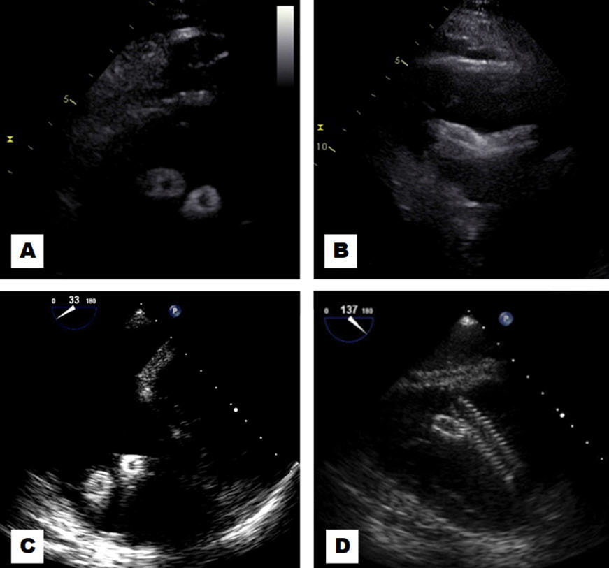
Lost vascular stents following endovascular repair of an arteriovenous dialysis graft were incidentally found peeking through the right ventricular (RV) inflow views on transthoracic (A) and transesophageal (C) echocardiography in short axis. Long axis transthoracic views (B) showed the stents crossing one another, and transesophageal (D) views showed the zipper-like appearance of the vascular stents at the RV inflow adjacent to the tricuspid valve. Vascular stents are increasingly utilized for endovascular interventions in dialysis grafts [1], including hybrid grafts [2]. This comes at an increased risk of acute and long-term complications such as stent fracture or migration [3]. Although a wait-and-watch approach is initially prudent, there are a number of bail-out interventional procedures available [4].
References:
Authors:
Landai Nguyen, D.O.
Cardiology Fellow
University of South Alabama
Mobile, AL
Sarina Sachdev, M.D.
Cardiology Fellow
University of South Alabama
Mobile, AL
Hassan Tahir, M.D.
Cardiology Fellow
University of South Alabama
Mobile, AL
Farnoosh Rahimi, M.D.
Assistant Professor of Cardiology
University of South Alabama
Mobile, AL
Barbara Burckhartt, M.D.
Associate Professor of Cardiology
University of South Alabama
Mobile, AL
Christopher Malozzi, D.O.
Assistant Professor of Cardiology
University of South Alabama
Mobile, AL
Bassam Omar, M.D., Ph.D.
Professor of Cardiology
University of South Alabama
Mobile, AL
G. Mustafa Awan, M.D.
Associate Professor of Cardiology
University of South Alabama
Mobile, AL
Lost vascular stents following endovascular repair of an arteriovenous dialysis graft were incidentally found peeking through the right ventricular (RV) inflow views on transthoracic (A) and transesophageal (C) echocardiography in short axis. Long axis transthoracic views (B) showed the stents crossing one another, and transesophageal (D) views showed the zipper-like appearance of the vascular stents at the RV inflow adjacent to the tricuspid valve. Vascular stents are increasingly utilized for endovascular interventions in dialysis grafts [1], including hybrid grafts [2]. This comes at an increased risk of acute and long-term complications such as stent fracture or migration [3]. Although a wait-and-watch approach is initially prudent, there are a number of bail-out interventional procedures available [4].
References:
- Shemesh D, Goldin I, Olsha O. Stent grafts for treatment of cannulation zone stenosis and arteriovenous graft venous anastomosis. J Vasc Access. 2017 Mar 6;18(Suppl. 1):47-52.
- Gomez LF, Peden EK. Description and early outcomes of the hybrid graft for dialysis. J Vasc Access. 2017 Mar 6;18(Suppl. 1):64-67.
- Ho JM, Kahan J, Supariwala A, et al. Vascular stent fracture and migration to pulmonary artery during arteriovenous shunt thrombectomy. J Vasc Access. 2013 Apr-Jun;14(2):175-9.
- Sequeira A. Stent migration and bail-out strategies. J Vasc Access. 2016 Sep 21;17(5):380-5.
Authors:
Landai Nguyen, D.O.
Cardiology Fellow
University of South Alabama
Mobile, AL
Sarina Sachdev, M.D.
Cardiology Fellow
University of South Alabama
Mobile, AL
Hassan Tahir, M.D.
Cardiology Fellow
University of South Alabama
Mobile, AL
Farnoosh Rahimi, M.D.
Assistant Professor of Cardiology
University of South Alabama
Mobile, AL
Barbara Burckhartt, M.D.
Associate Professor of Cardiology
University of South Alabama
Mobile, AL
Christopher Malozzi, D.O.
Assistant Professor of Cardiology
University of South Alabama
Mobile, AL
Bassam Omar, M.D., Ph.D.
Professor of Cardiology
University of South Alabama
Mobile, AL
G. Mustafa Awan, M.D.
Associate Professor of Cardiology
University of South Alabama
Mobile, AL
Challenging Images
Tumors Uptake Microbubbles..Gee Fizz!
Description
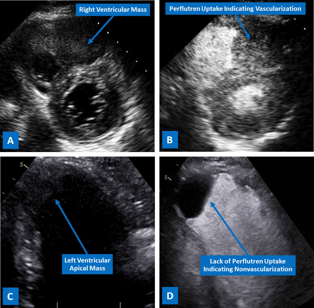
Intracardiac masses not only can be poorly visualized by transthoracic echocardiography, but can also be difficult to characterize based on their appearance only [1]. In the figure, the mass seen within the right ventricle on the parasternal short axis view of a 2-D echocardiogram (A) readily uptakes perflutren microbubbles (Definity), indicting its vascularity (tumor). In contrast, the left ventricular mass (C) seen on the apical 2-chamber view does not uptake microbubbles (D) indicating lack of vascularity (thrombus). Contrast echo plays an essential role in endocardial border delineation [2], in addition to characterization of cardiac masses [3], especially in difficult to image patients [4].
References:
Authors:
Landai Nguyen, D.O.
Cardiology Fellow
University of South Alabama
Mobile, AL
Sarina Sachdev, M.D.
Cardiology Fellow
University of South Alabama
Mobile, AL
Sajjad Ahmad, M.D.
Cardiology Fellow
University of South Alabama
Mobile, AL
Farnoosh Rahimi, M.D.
Assistant Professor of Cardiology
University of South Alabama
Mobile, AL
Barbara Burckhartt, M.D.
Associate Professor of Cardiology
University of South Alabama
Mobile, AL
Christopher Malozzi, D.O.
Assistant Professor of Cardiology
University of South Alabama
Mobile, AL
Bassam Omar, M.D., Ph.D.
Professor of Cardiology
University of South Alabama
Mobile, AL
Intracardiac masses not only can be poorly visualized by transthoracic echocardiography, but can also be difficult to characterize based on their appearance only [1]. In the figure, the mass seen within the right ventricle on the parasternal short axis view of a 2-D echocardiogram (A) readily uptakes perflutren microbubbles (Definity), indicting its vascularity (tumor). In contrast, the left ventricular mass (C) seen on the apical 2-chamber view does not uptake microbubbles (D) indicating lack of vascularity (thrombus). Contrast echo plays an essential role in endocardial border delineation [2], in addition to characterization of cardiac masses [3], especially in difficult to image patients [4].
References:
- Mankad R, Herrmann J. Cardiac tumors: echo assessment. Echo Res Pract. 2016 Dec;3(4):R65-R77..
- Kaufmann BA, Wei K, Lindner JR. Contrast echocardiography. Curr Probl Cardiol. 2007 Feb;32(2):51-96.
- Kirkpatrick JN, Wong T, Bednarz JE, et al. Differential diagnosis of cardiac masses using contrast echocardiographic perfusion imaging. J Am Coll Cardiol. 2004 Apr 21;43(8):1412-9.
- Daniel GK, Chawla MK, Sawada SG, et al. Echocardiographic imaging of technically difficult patients in the intensive care unit: use of optison in combination with fundamental and harmonic imaging. J Am Soc Echocardiogr. 2001 Sep;14(9):917-20.
Authors:
Landai Nguyen, D.O.
Cardiology Fellow
University of South Alabama
Mobile, AL
Sarina Sachdev, M.D.
Cardiology Fellow
University of South Alabama
Mobile, AL
Sajjad Ahmad, M.D.
Cardiology Fellow
University of South Alabama
Mobile, AL
Farnoosh Rahimi, M.D.
Assistant Professor of Cardiology
University of South Alabama
Mobile, AL
Barbara Burckhartt, M.D.
Associate Professor of Cardiology
University of South Alabama
Mobile, AL
Christopher Malozzi, D.O.
Assistant Professor of Cardiology
University of South Alabama
Mobile, AL
Bassam Omar, M.D., Ph.D.
Professor of Cardiology
University of South Alabama
Mobile, AL


